Cross Sections of the Spinal Cord
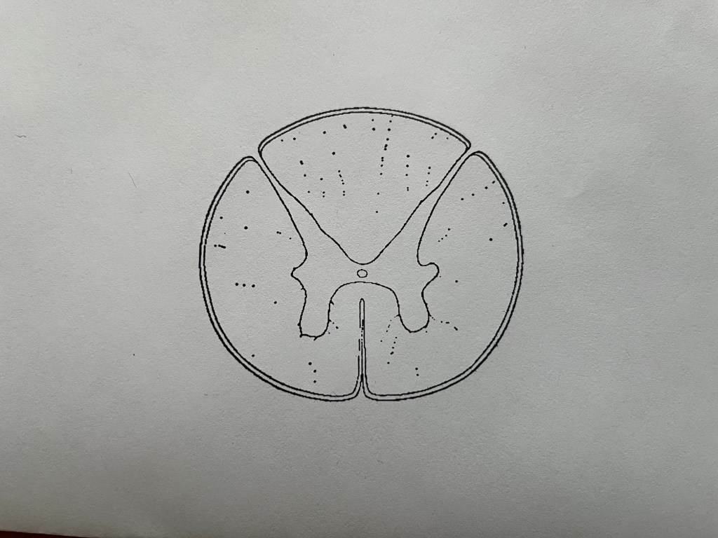
This is a drawing of s section of the spinal cord, a central nervous system structure. The top of the image is the back of the spinal cord, called dorsal (Latin for back). The bottom of the image is the front, or ventral aspect of the spinal cord. The butterfly looking structure in the center is the gray matter, meaning that it consists of neuron cell bodies, which look gray in dissection. The top wings, or horns as they are called, are the dorsal horns and the ones below are the ventral horns. Remember, in the nervous system, motor signals come out the front and sensory comes in the back. The ventral horns contain motor neurons which are called lower motor neurons and also called alpha motor neurons. They say that anatomists were paid by the name, so multiple structures have multiple names, often for good reason. We will get to these distinctions later when we discuss proprioception. The dorsal horns contain sensory neurons, also known as second order sensory neurons. The first order sensory neurons are in the peripheral nervous system, as you will see below.
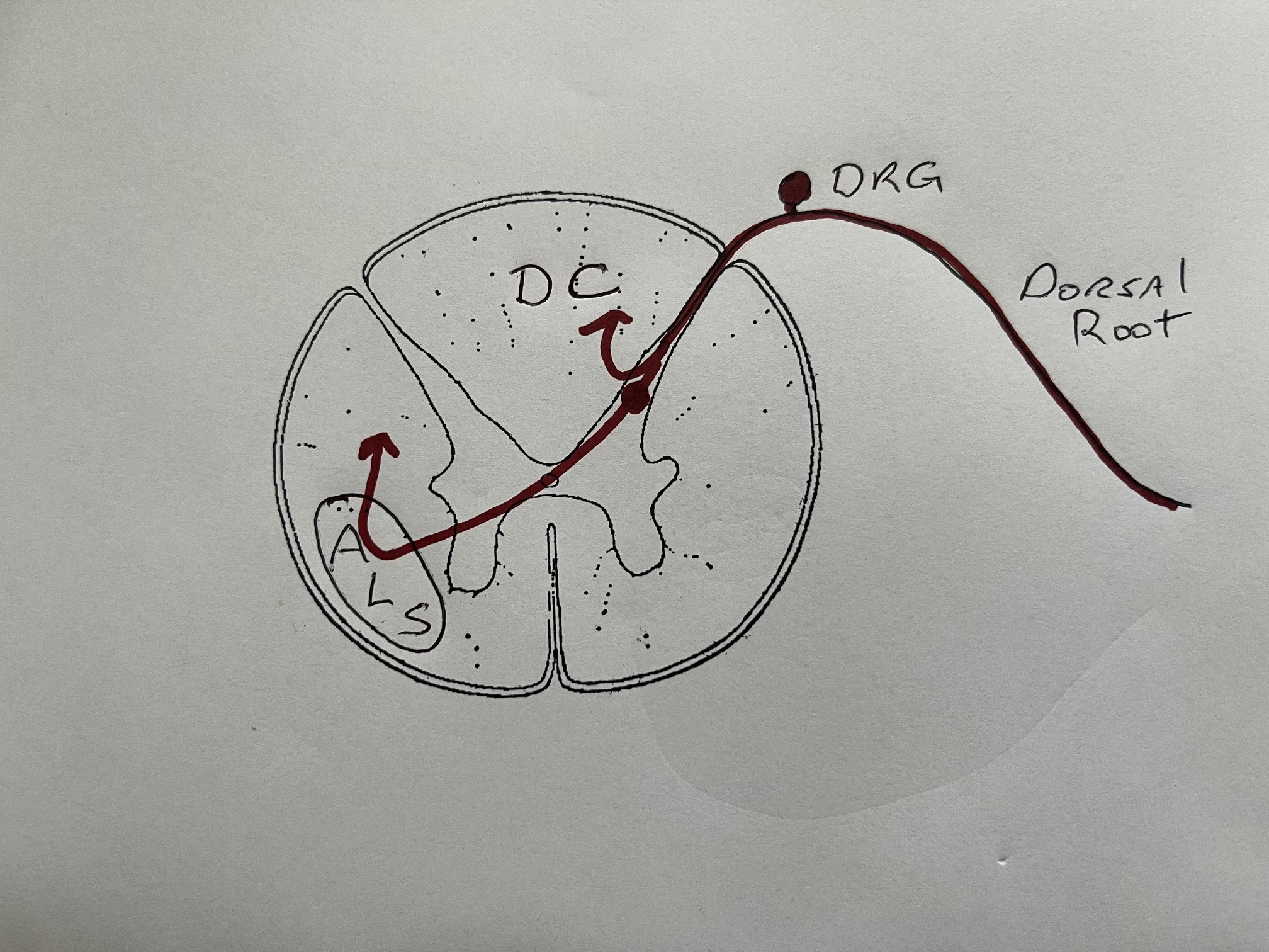
Let’s look at the dorsal root of the spinal cord first. Sensory information from sensory receptors on and in the body travels up sensory nerve axons and enters the dorsal horn of the spinal cord. The first order sensory neuron cell body is in the dorsal root ganglion, which lies outside the dura, in the peripheral nervous system. There is no synapse in this ganglion, only a sensory neuron cell body. The term ganglion is a problem in neuroanatomy, as it means different things depending on what functions you are talking about. Somatosensory ganglia do not contain a synapse. Visceromotor ganglia have synapsing neurons. Soma means body in Greek. Again, with all the names.
That somatosensory information, body sensory information as opposed to sensory information from the guts, called viscerosensory. The somatosensory information is of two types and takes two separate routes to the brain. Knowing the separate routes is essential to localizing lesions in patients. The first type of somatosensory information is called fine touch and proprioception. This information peels off medially (toward the center) at it enters the dorsal horn, and the axons ascend the spinal cord toward the brainstem in a set of columns, called the dorsal columns (DC). The second type of somatosensory information is pain and temperature. This information enters the dorsal horn and synapses on second order neurons in the dorsal horn, and then those axons (secondary sensory) cross to the other side of the spinal cord and ascend toward the midbrain in a bundle of axons called the anterolateral system (ALS). This is important in diagnosing lesions as some symptoms will be on the same side of the body as the injury, and some will be on the other.
Fine touch is the sense that allows you to discern exactly where you are being touched, depending on the density of the receptors. You have more fine touch receptors on your fingers than you do on your back. Proprioception is the sense of where your body is in three-dimensional space, and in what direction you are moving and how fast. Proprioception is essential to balance and movement and disruptions can severe.
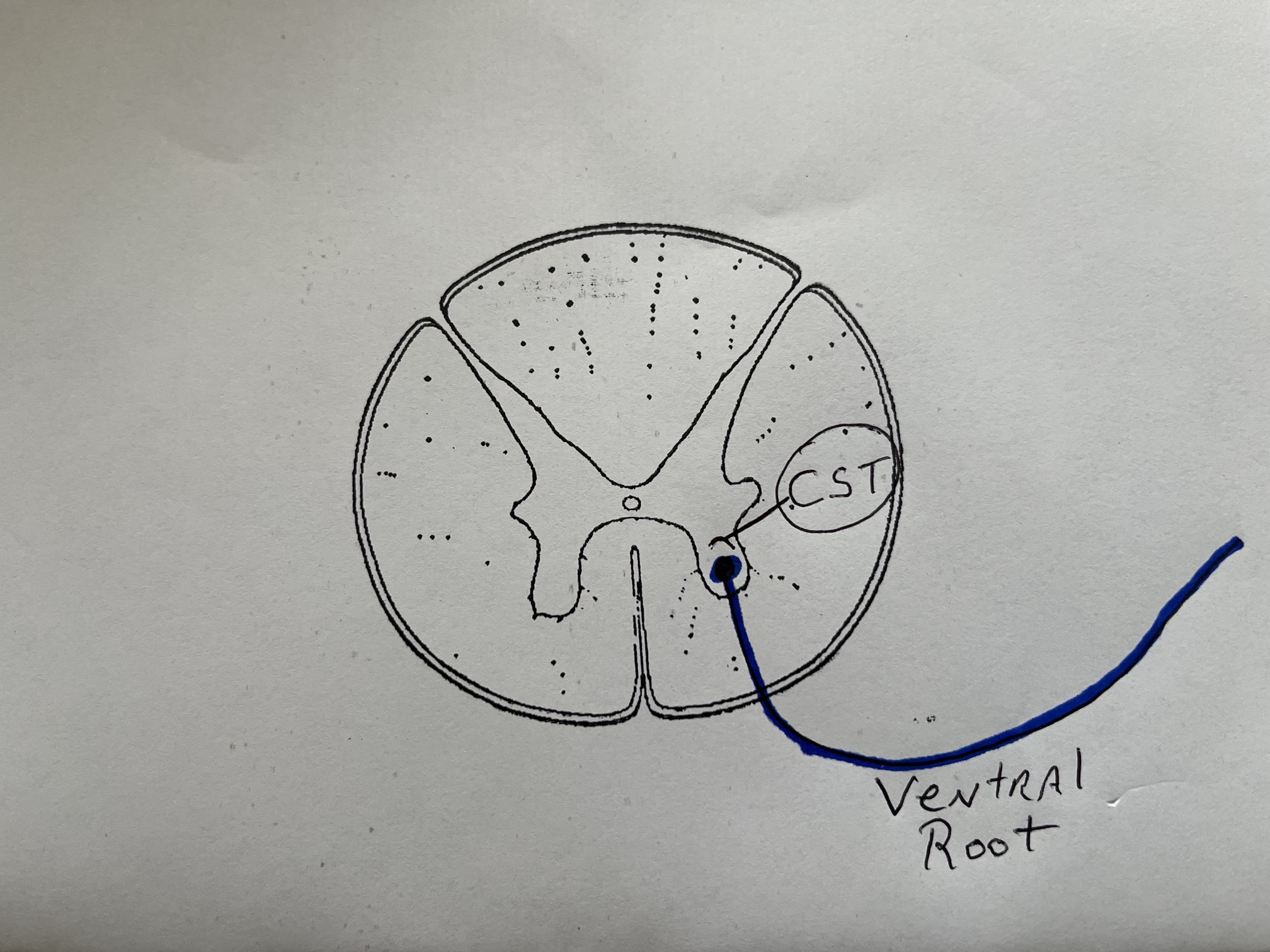
This image shows the ventral root of the spinal nerve. As you know, motor comes out the front, so this is a motor nerve. Remember that there is one on each side. The alpha motor neuron cell body is in the ventral horn. Motor signals from the frontal lobe of the brain come down the spinal cord in a bundle called the corticospinal tract (CST). Those neuron cell bodies in the frontal lobe are called upper motor neurons. Damage to upper motor neurons causes a rigid type of paralysis called spastic paralysis. Damage to lower motor neurons, or their axons, causes flaccid paralysis. The axon of the lower motor neurons exits the cord as the ventral root and are then routed to skeletal muscles along that spinal level.
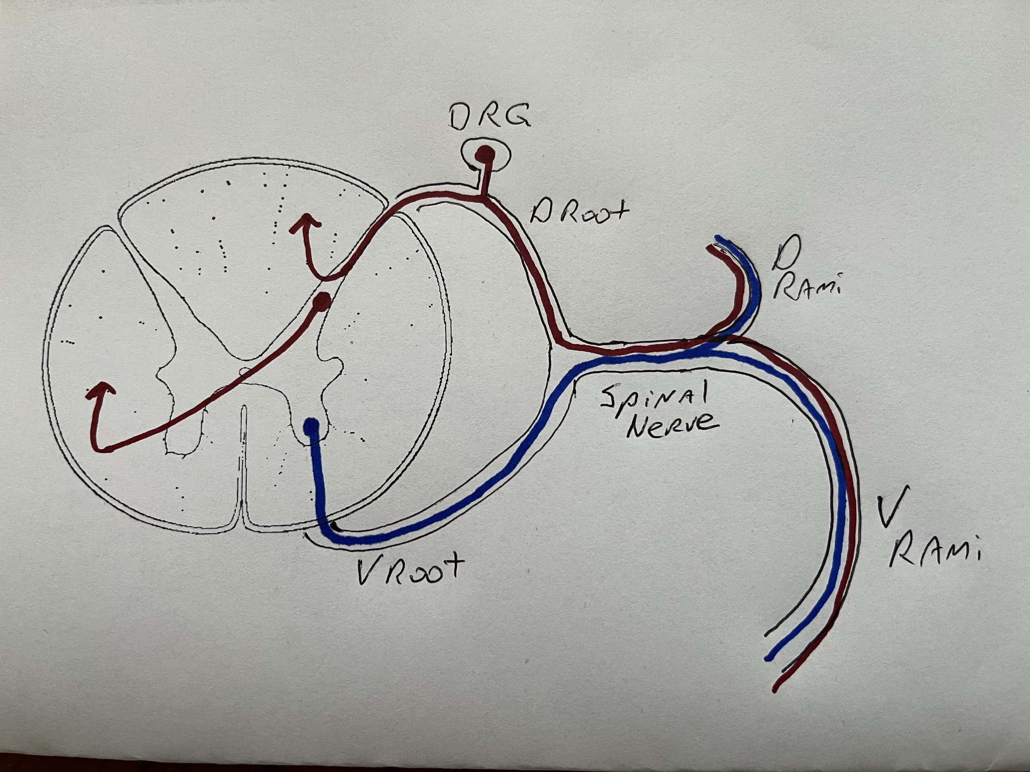
Now let’s put it together. The spinal nerve, one on each side of the spine, comes out of a segment of the spinal cord named for the numbered vertebra in your spine. You have 31 pairs of spinal nerves named for their region of origin – cervical (C1-C8), thoracic (T1-T12), lumber (L1-L5), sacral (S1-S5), and a single coccygeal pair. Each spinal nerve innervates a specific area called a dermatome.
Spinal nerves have roots, coming in and out of the spinal cord, and rami, which are branches. The is a large ventral ramus going to the sides and front of the body, and a smaller dorsal ramus going to the back. As you can see, the roots contain only sensory or motor axons, while the rami contain both sensory and motor coming from and going to the body.
 Return Human Nervous System Home Page