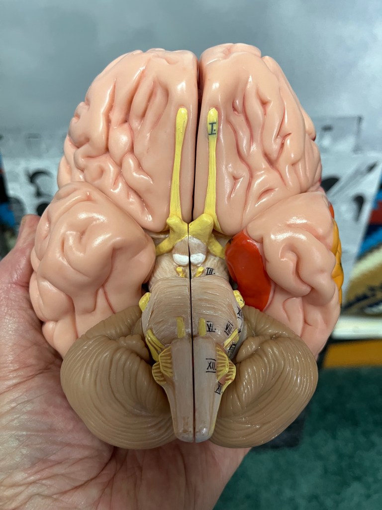
Starting at the top of the image, you can see the two long olfactory tracts coming from cranial nerve I, the olfactory nerves. Remember that there is one on each side of the brain or brainstem. The olfactory tracts lie on the inferior surface of the frontal lobes and send olfactory information to the frontal lobes and the temporal lobes, specifically the amygdala.
Moving toward the center of the image, you see the optic chiasm, which are the optic nerves, cranial nerves II, crossing to go to the contralateral side of the brain. The axons from retinal (ganglion) cells on the temporal side stay ipsilateral and the ganglion cells from the nasal retina cross to the other side, such that the right visual field goes to the left side of the thalamus and the occipital lobes, and the left visual filed goes to the right side of the thalamus and the occipital lobes.
Moving caudally, the next nerves, cranial nerves III, are the oculomotor nerves, which move most of the muscles of the ipsilateral eyes. It is hard to see, but cranial nerves IV, the trochlear nerves are wrapping around from the back side of the midbrain/pons junction to power the superior obliques in the eye, which intort the eyes when you are walking and looking down the stairs.
Cranial nerves V, the trigeminal nerves are the large bulbs sitting on the lateral aspects of the pons. They receive sensory information from the face and power the jaw muscles for chewing.
Moving down in the image, you can see the pairs of nerves from the center to the periphery, coming out of the junction between the pons and the medulla. These are cranial nerves VI (center-most), VII, and VIII. CN VI, the abducens nerves, abduct the ipsilateral eyes; CN VII, the facial nerves, move the facial muscles used in emotional expression and receives taste information from the front 2/3rd of the tongue; and CN VIII, the vestibulocochlear nerves, conduct the senses of hearing and balance.
Below that, the most medial nerves on the medulla, are the hypoglossal nerves, CN XII, which power the tongue. There is a little bump, called the olive, just posterior to the CNs XII. Behind the olive, CNs IX, glossopharyngeal, X, vagus, and XI accessory, form a spider web that exits the jugular foramen heading for the trapezius and SCL muscles (XI), and the throat, esophagus (IX), lungs, heart, and guts (X).
For a more detailed explanation of the cranial nerves, see the chapter in my book Understanding the Big Picture of the Nervous System: Sorrells PhD, Robert: 9798307936429: Amazon.com: Books
