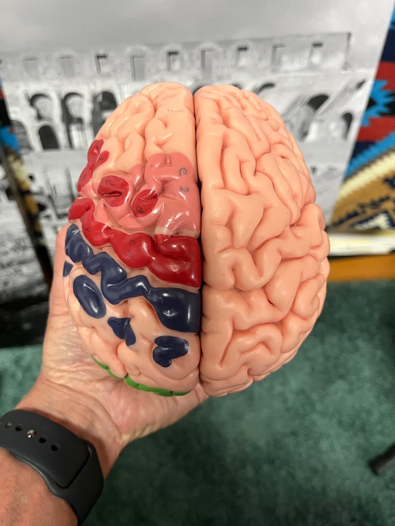
Looking down onto the top of the brain. You can see that the left and right sides, the hemispheres, look nearly identical. The left side of the brain controls the right side of the body and vice-versa. Sensory input to the brain also comes from the other side, the contralateral side
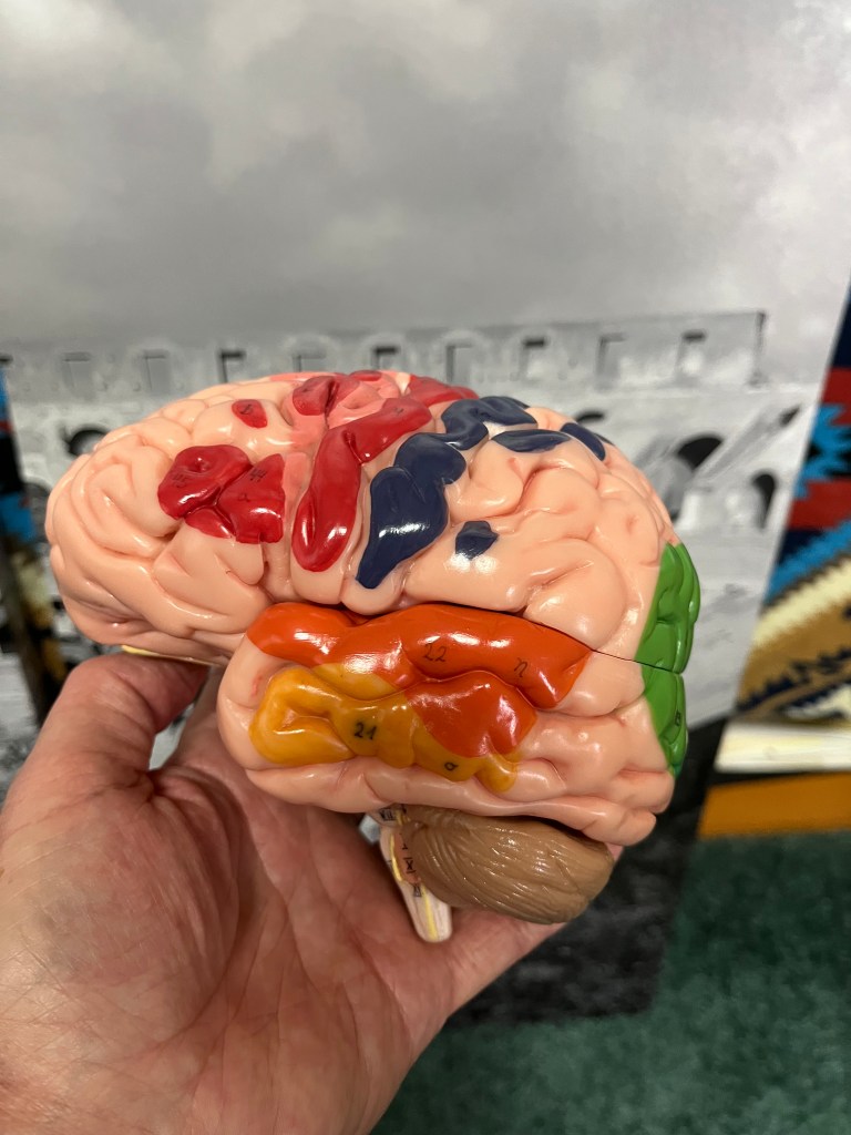
Here is a look at the left hemisphere of the brain. The grooves are called sulci (sulcus) and the ridges are called gyri (gyrus). The groove between the red and blue gyri is the central sulcus and marks the division between the motor cortex in the front, and the sensory cortex in the back.
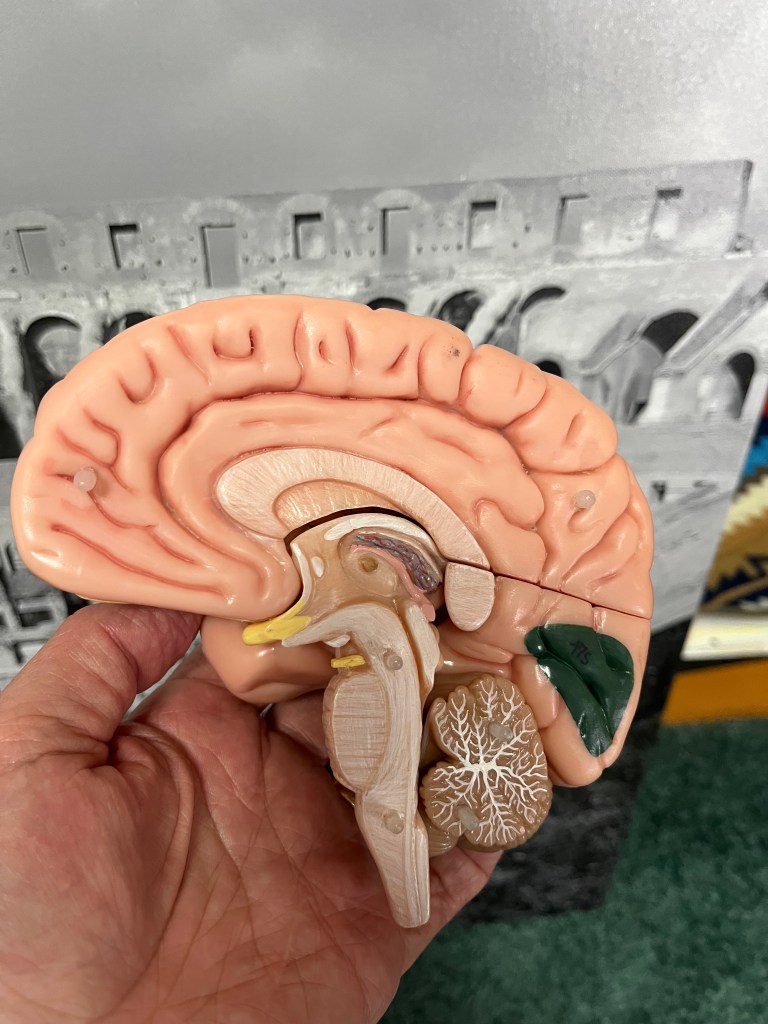
Here, the model is taken apart into left and right sides. This is the inside of the right half. This view is called mid sagittal in imaging and dissection. You can see the medulla at the bottom of the brain stem, and the bulge above it is the pons, and the midbrain sits on top of the pons. The cerebellum, which ensures coordination, is hanging off the back. Those white spidery lines are bundles of axons, white matter. The third ventricle and the thalamus are right in the center, capped by the corpus collosum, which is a huge (colossal) band of axons going back and forth between the hemispheres. The cingulate gyrus sits above the corpus callosum and produces emotional reactions, decision making, and memory.
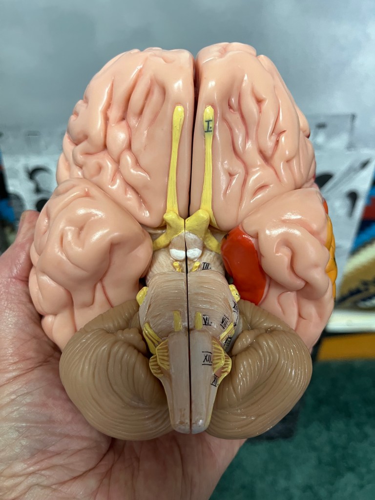
This is a view from the bottom, a ventral view. The brainstem is prominent, and you can easily see the hypoglossal nerves, cranial nerve XII, which are the only cranial nerves to exit the medulla in from of a bump called the olive. Cranial nerves VI, VII, and VIII are coming out of the depression between the medulla and pons. The large crossing object is the optic chiasm, and the two long nerves on the floor of the frontal lobes are the olfactory nerves. I’ll spend more time going over these structures as the webpage continues to grow.
