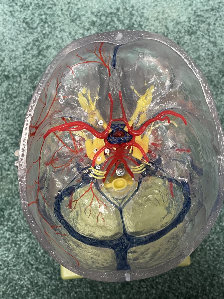
Looking down into the skull with the brain removed you can see the arterial supply and most of the venous draining from the brain. In the bottom of the image, you can see the confluence of sinuses. This is the junction of the superior sagittal sinus coming from the top of the brain, the straight sinus draining the middle of the brain, and the occipital sinuses. Venous blood will drain left or right along the transverse sinuses, down the sigmoid sinuses and into the internal jugular veins.
At the top of the image, you can see the blue cavernous sinuses surrounding the pituitary stalk. Surrounding that is the Circle of Willis, the hub of brain blood flow. The Circle of Willis, and most of this blood supple would be stuck to the bottom of the brain by a layer a pia, but it is easier to see like this. You can see the vertebral arteries like two legs joining together to become the basilar artery, which together irrigate the brainstem and cerebellum, and continue to ascend and then split into the posterior cerebral arteries, which turn back and head for the occipital lobes. Branching off each posterior cerebral artery is a posterior communicating artery joining the circle. The next two large arteries you see are the left and right middle cerebral arteries, which come off the internal carotids as they ascend into the skull. The middle cerebral arteries spill out onto the lateral surface of the cerebral hemispheres are common sites for aneurisms and stroke. You also see the smaller left and right anterior cerebral arteries moving forward to supply cortex on the midsagittal aspect of each hemisphere, down in the longitudinal fissure. They are connected by a small but important branch, the anterior communicating artery. The optic chiasm is underneath and can be damaged by berry aneurisms on the anterior communicating artery. Pituitary tumors can also press onto the optic chiasm causing bitemporal hemianopias, AKA tunnel vision.
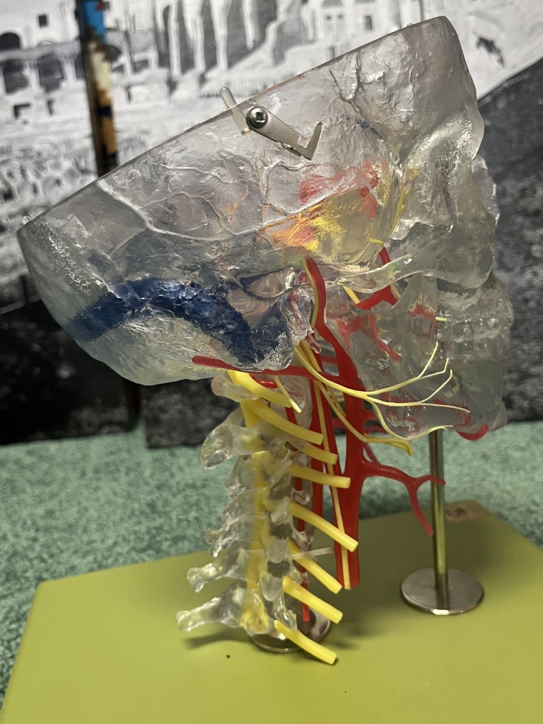
Here is side view of Skully. You can see the vertebral processes sticking back from the cervical vertebral column. The largest on the bottom is the vertebral prominens of C7 and can easily be felt on the back of your neck. Count up from there to C2, Axis, and C1 stuck to the bottom of the skull, Atlas. You can see the spinal cord, in yellow, inside the vertebral column, and the large spinal nerves coming out. In front of all that is the larger right carotid artery, or the common carotid artery. It splits into the external carotid artery moving forward, supplying the thyroid, face, and mouth, and the internal carotid artery climbing up into the skull to join the Circle of Willis.
